Foot & Ankle Medical Imaging

Podiatrists will often refer patients to have an x-ray, ultrasound and/or MRI of the foot and ankle to assist in the diagnosis and management of certain pathologies.
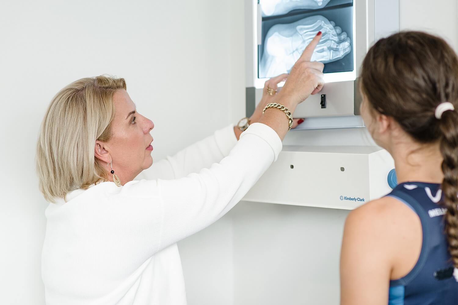
Podiatrists are skilled at reading and interpreting foot and ankle medical imaging and this can be of help in the confirmation of pathology, assessing structural alignment, assessment of inflammation and treatment planning for patients.
Medical imaging is something podiatrists can refer for to assist in understanding baselining pathology.
X-rays of the foot and ankle is a two-dimensional image of the body’s bones to help diagnose conditions or disease, similar to a photograph of your bones. An x-ray can be used to assess alignment of the foot and ankle whilst sitting or standing. Diagnosis of certain bony pathology include stress fractures, bony infection and bunions to name a few. This can be covered by Medicare and occasionally may have a gap fee associated with it.
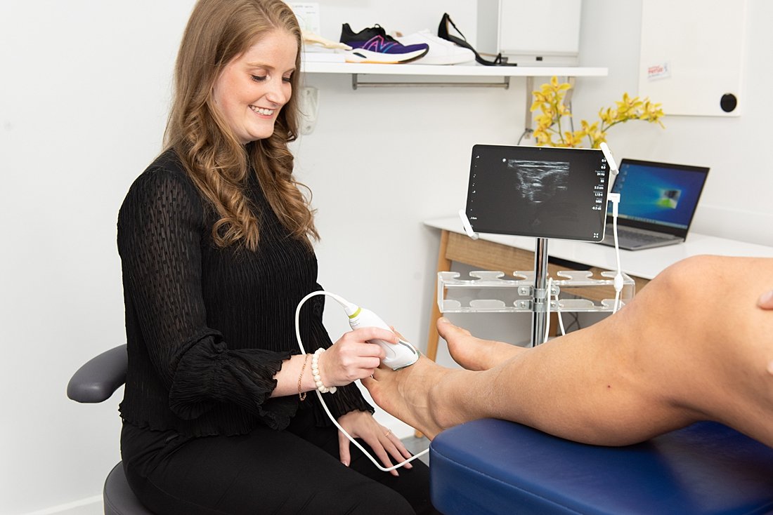
Ultrasound of the foot and ankle can be ordered to allow the podiatrist to assess tendon pathology including tears, ligament issues, muscle patholody, nerve problems and inflammation of certain areas of the foot and ankle. An ultrasound is a non invasive technique that uses high frequency sound waves to create an image of the foot and ankle.
An MRI (Magnetic Resonance Imaging) is a noninvasive nuclear procedure for imaging tissues of high fat and water content that cannot be seen with other radiologic techniques. The MRI image gives information about the chemical makeup of tissues, thus making it possible to distinguish normal, atherosclerotic, and traumatised tissue masses in the image. An MRI can be ordered by podiatrists, however they do attract a fee the same as if your GP ordered one for you – MRIs are only subsided by specialist doctors. MRIs are beneficial to determine soft tissue and bony pathology that aren’t clearly identified on ultrasound or x-ray.

Sophie Young, Rebecca Neaves, and Kirstine Mann have undergone further training to use our in-clinic diagnostic ultrasound. They can use this on patients to diagnose and assess soft tissue pathology. The in-clinic ultrasound also enables corticosteroid injections under ultrasound guidance. Sophie and Rebecca can also administer corticosteroid injections in the clinic as they have completed their 150 practical hours as part of her Endorsement to Schedule Medicine studies.
Always Consult A Trained Professional
The information in this resource is general in nature and is only intended to provide a summary of the subject matter covered. It is not a substitute for medical advice and you should always consult a trained professional practising in the area of medicine in relation to any injury or condition. You use or rely on information in this resource at your own risk and no party involved in the production of this resource accepts any responsibility for the information contained within it or your use of that information.
